Experience the Intense Challenge and Deep Reward of Dark Souls III 3 Deluxe Edition PC
Hi everyone! Are you familiar with Dark Souls III 3 Deluxe Edition PC? It’s the most recent entry in the well-liked action role-playing game, Dark Souls. I can’t wait to tell you everything about it! The Dark Souls games are known for being super challenging but also rewarding. They don’t hold your hand at all, the combat is strategic and punishing, and the levels are intricately designed. But man, when you finally beat a tough boss or make it through a treacherous area, the sense of accomplishment is awesome!
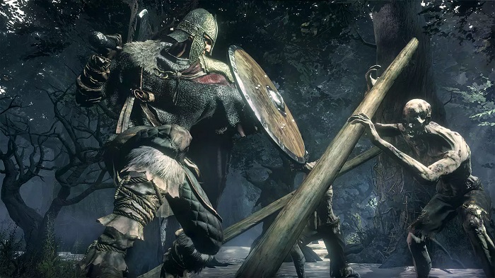
You make your unique avatar in Dark Souls III and embark on an adventurous quest across the realm of Lothric. To prevent the planet from falling into complete darkness, you must rekindle the First Flame. However, the trip will not be easy. Your reflexes and thinking will be tested in Lothric’s many difficult boss encounters, lethal traps, and formidable opponents.
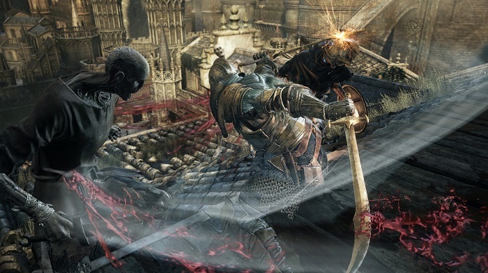
The combat in Dark Souls III deluxe edition download code builds on the franchise’s signature strategic real-time combat. You must carefully time your dodges, look for enemy attack patterns, manage your stamina, and strike at the right moments. Each weapon has its moveset that you’ll need to master, too. And you can equip different weapons and armor to match your playstyle.
Mastering Dark Souls III 3 Deluxe Edition PC Strategic Combat and Rich RPG Customization Offers
The role-playing game components are developed further in the third game. You can level up, boost your health and stamina, and gain new skills as you play. It’s important to balance offensive, defensive, and utility skills. There are also many magical spells, armor sets, swords, and other pieces of equipment to unearth.
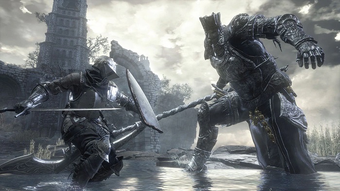
The boss battles are still awesome, of course! We are discussing corrupted creatures, dragons, knights, and enormous demons. They all have intricate assault patterns you must become familiar with and avoid. It requires perseverance, talent, and precise tactics to defeat these bosses. But after several tries, it’s so satisfying when you eventually defeat one.
The World of PC Dark Souls iii 3 deluxe edition is intricately designed with shortcuts, hidden secrets, and deadly traps. You’ll need to pay close attention to your surroundings at all times. Optional areas are sprinkled throughout, too, rewarding exploration. And you can play online to cooperate or compete with other players for that extra challenge. So, that’s a quick summary of what Dark Souls III has to give. You should try it if you want a tough, strategic combat task with rewarding gameplay. Know that it won’t be simple. But it’s worth it when you finally beat a boss or find a secret spot. If you have any more questions, please let me know.
Characters in Dark Souls III 3 Deluxe Edition PC
- The Ashen One: This is the player character whose fate is uncertain. As an Unkindled, unfit even to be cinder, you have been resurrected to link the First Flame and prevent an Age of Dark.
- Ludleth of Courland: A Lord of Cinder who remains in Firelink Shrine. He provides key insights into the world’s mythos and helps the Ashen One on their journey.
- Fire Keeper: This quiet and mystical woman tends to the bonfire at Firelink Shrine. She assists the Ashen One by providing leveling-up services.
- Hawkeye Gough: A giant and legendary archer who expounds lore and helps take down a formidable dragon. His skills with a bow are unmatched.
- Siegward of Catarina: A bumbling knight in onion-shaped armor who repeatedly helps the player in jolly cooperation. He enjoys his ale and sticking to his oaths.
- Anri of Astora: A kind, Unkindled undead traveling on their quest who crosses paths with the Ashen One several times. Their fates are intertwined.
- Yuria of Londor: A mysterious woman who serves the primordial serpent Kaathe and tries to guide the Ashen One down the path of the Lord of Hollows.
- Pontiff Sulyvahn: A cruel sorcerer and self-proclaimed high priest who has usurped control of Irithyll. He stands as a difficult mid-game boss.
- Dragonslayer Armour: This enchanted suit of armor defends the Grand Archives from intruders. The armor seems to move and fight off its own will.
- Sister Friede: A rogue sorceress who has usurped the Painted World of Ariandel for her selfish designs. She and Father Ariandel are difficult late-game bosses.
- Soul of Cinder: The deific manifestation of the previous Lords of Cinder. This amalgamation of their souls is the final challenge guarding the First Flame.
The most popular and useful mods that exist for Dark Souls III 3 Deluxe Edition PC
- Cinders Mod: This massive overhaul mod aims to provide a fresh experience. It adds new items, spells, weapons, bosses, etc., while expanding lore and content. Makes the game more challenging.
- Champion’s Ashes: Adds over 100 new combat animations and moves to expand the combat options. Lets you create more dynamic and customized playstyles.
- Convergence Mod: Includes new questlines, bosses, weapons, spells, and armor to discover. Also tweaks gameplay and systems for rebalancing.
- Arena Overhaul: Revamp the PvP arena with new maps, spectator tools, and quality-of-life changes like elastic matchmaking.
- Blue Sentinels Mod: Adds anti-cheat and anti-hacker protections to prevent malicious players from ruining the online experience.
- PKGI Mod: You can turn normal weapons into the ultra-rare Twinkling and Demon Titanite upgrade paths for greater build diversity.
- Randomizer Mod: Shuffles in-game items, enemy, and boss locations to create unpredictable new challenges on each playthrough. Keeps things fresh.
- First-Person Mode: Allows you to experience the entire game from a visceral, first-person perspective, increasing immersion and difficulty.
- Boss Rush Mode: Adds a gauntlet mode where you fight back-to-back bosses without breaks to test your skill. Leaderboards allow you to compete.
- Daughters of Ash Remastered: A massive fan-made overhaul adding new storylines, enemies, weapons, secrets, and mystique to Lordran.
updates that have been released for Dark Souls III 3 Deluxe Edition PC
- Launch Day Patch: This first patch fixed several game crashing bugs, balance issues, and exploits present at launch. Improved overall stability.
- Patch 1.01 to 1.12: Over the first year, these patches focused on incremental bug fixes, stability improvements, and small balance tweaks.
- Ashes of Ariandel DLC: Added the large new snowy area of the Painted World of Ariandel. Introduced new weapons, spells, bosses, and more.
- The Ringed City DLC: The final DLC added the dystopian city at the world’s end. Added epic new boss fights and a conclusive ending.
- Patch 1.13 to 1.15: Continued stability and performance improvements, particularly for the DLC areas. Fixed more exploits.
- 60 FPS Unlock: An update unlocked the framerate on the PC from the original 30 FPS cap, allowing for higher frame rates.
- Dark Souls III The Fire Fades Edition: This complete edition bundled the base game and DLC into one definitive edition.
- Covenant Rework Patch: Adjusted covenants like Blade of the Darkmoon for improved online activity and balance.
- Anticheat Patch: Improved cheat detections and banned invalid game data to improve online play integrity.
- AoA/TRC Weapon Matchmaking Fix: Prevented DLC weapons from matching with non-DLC owners to improve online connections.
- Blue Sentinels Anti-Cheat: Integrated the popular third-party Blue Sentinels anti-cheat mod natively into the game.
Minimum System Requirements
- OS: Windows 7 SP1 64bit, Windows 8.1 64bit, Windows 10 64bit
- Processor: Intel Core i3-2100 3.1GHz or AMD FX-6300 3.5GHz
- Memory: 8 GB RAM
- Graphics: NVIDIA GeForce GTX 750 Ti or AMD Radeon HD 7850 2GB
- DirectX: Version 11
- Storage: 50 GB available space
- Additional Notes: Xbox 360 Controller or DualShock 4 Controller recommended.